How We Hear Hearing Associates, Inc.
Download a free printable outline of this video and draw along with us: https://artforall.me/video/how-to-draw-human-earThank you for watching. Please subsc.
Hearing Sense Ask A Biologist
However, interactive ear diagrams change the game. Platforms like ESL Games Plus have introduced an exciting ear diagram to label, which allows students to drag and drop names of the ear's parts to their correct positions. This kind of interactive learning ensures better retention and understanding of the subject matter.
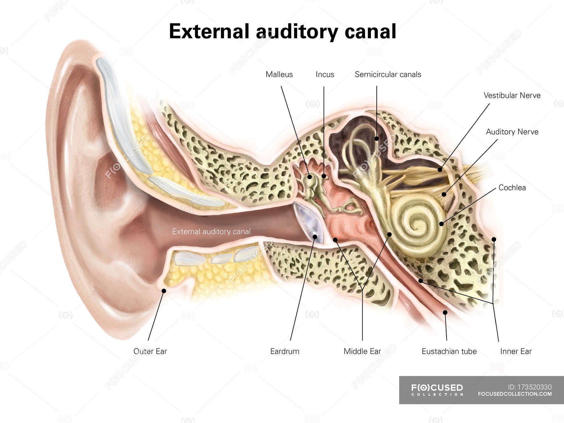
Auditory canal of human ear — vestibular, labels Stock Photo 173520330
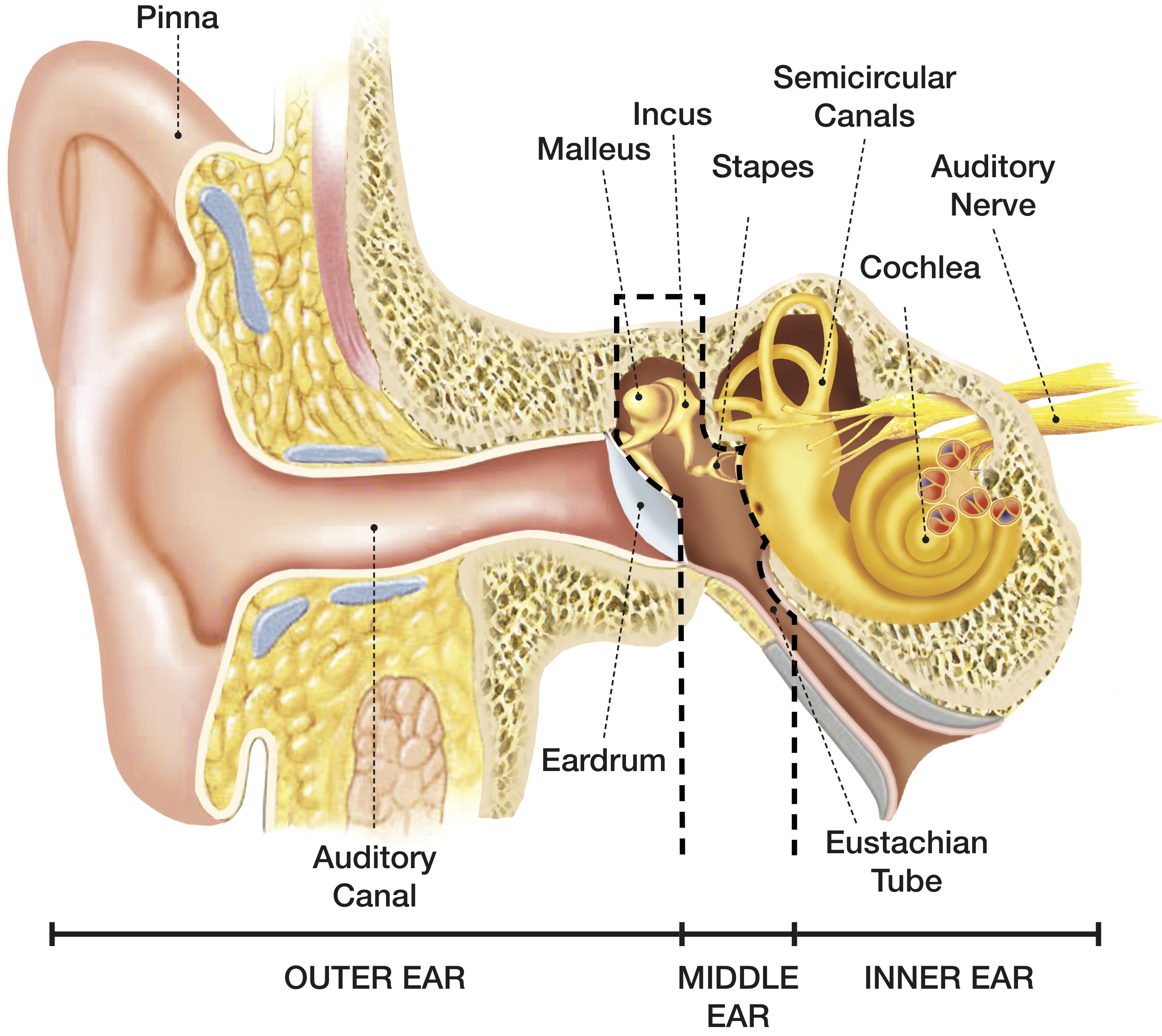
1: Diagram showing the structure of the human ear, detailing the parts of the outer, middle, and inner ear. Source publication +48 A Framework for Speechreading Acquisition Tools Thesis.
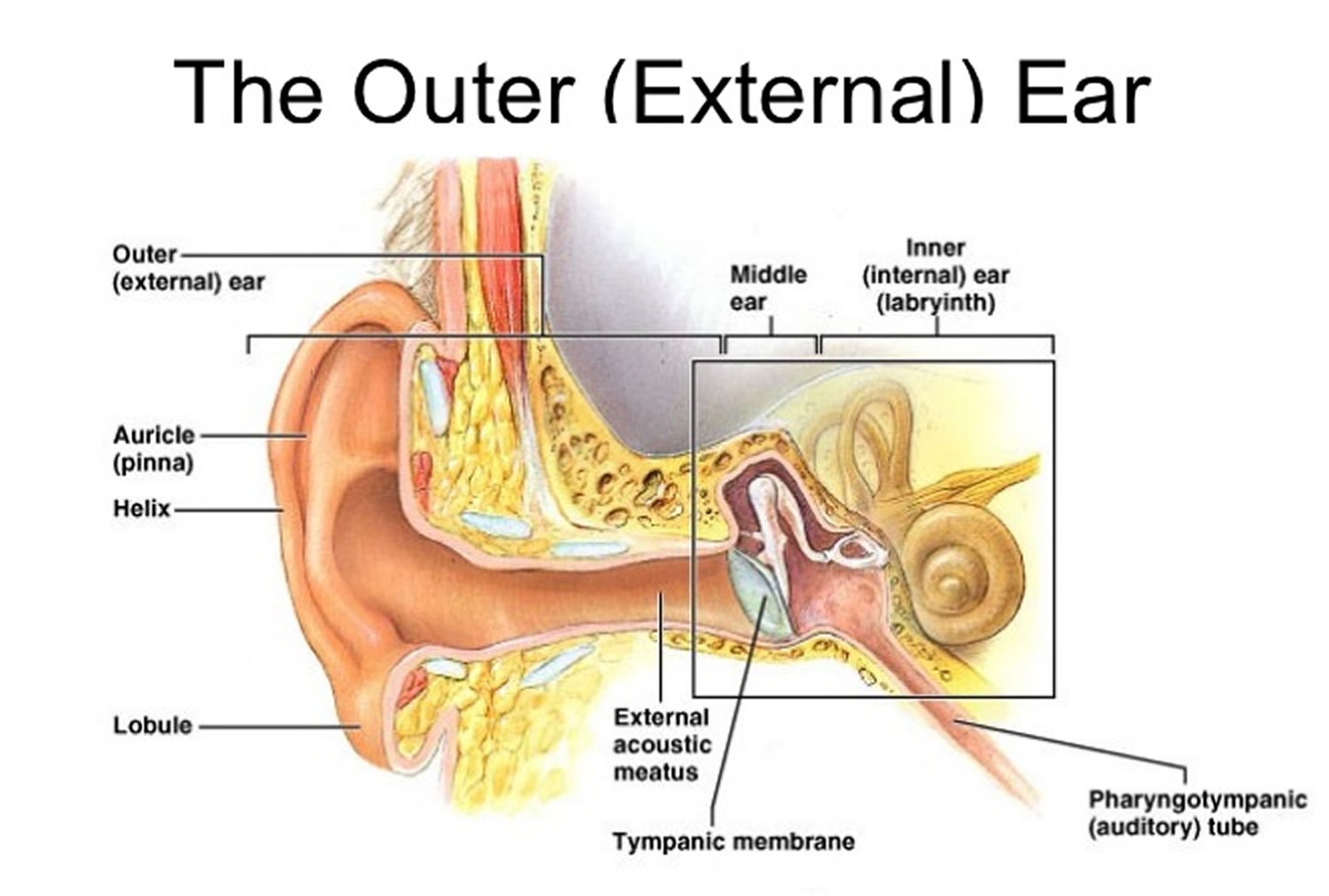
Outer Ear Anatomy Outer Ear Infection & Pain Causes & Treatment
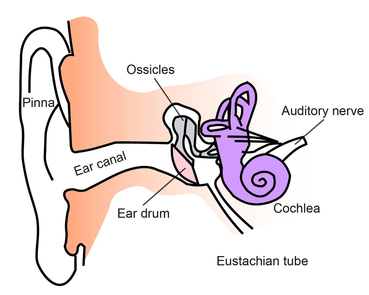
The following ear diagram depicts the inner ear, which contains sensory organs for hearing and balance, and the outer ear, which includes superficial structures.

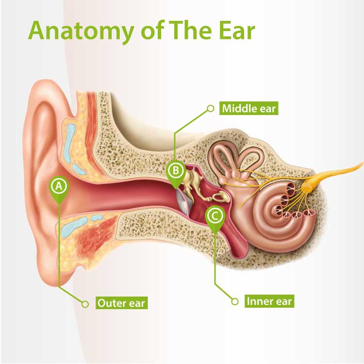
Anatomy of the Ear [4]. Download Scientific Diagram
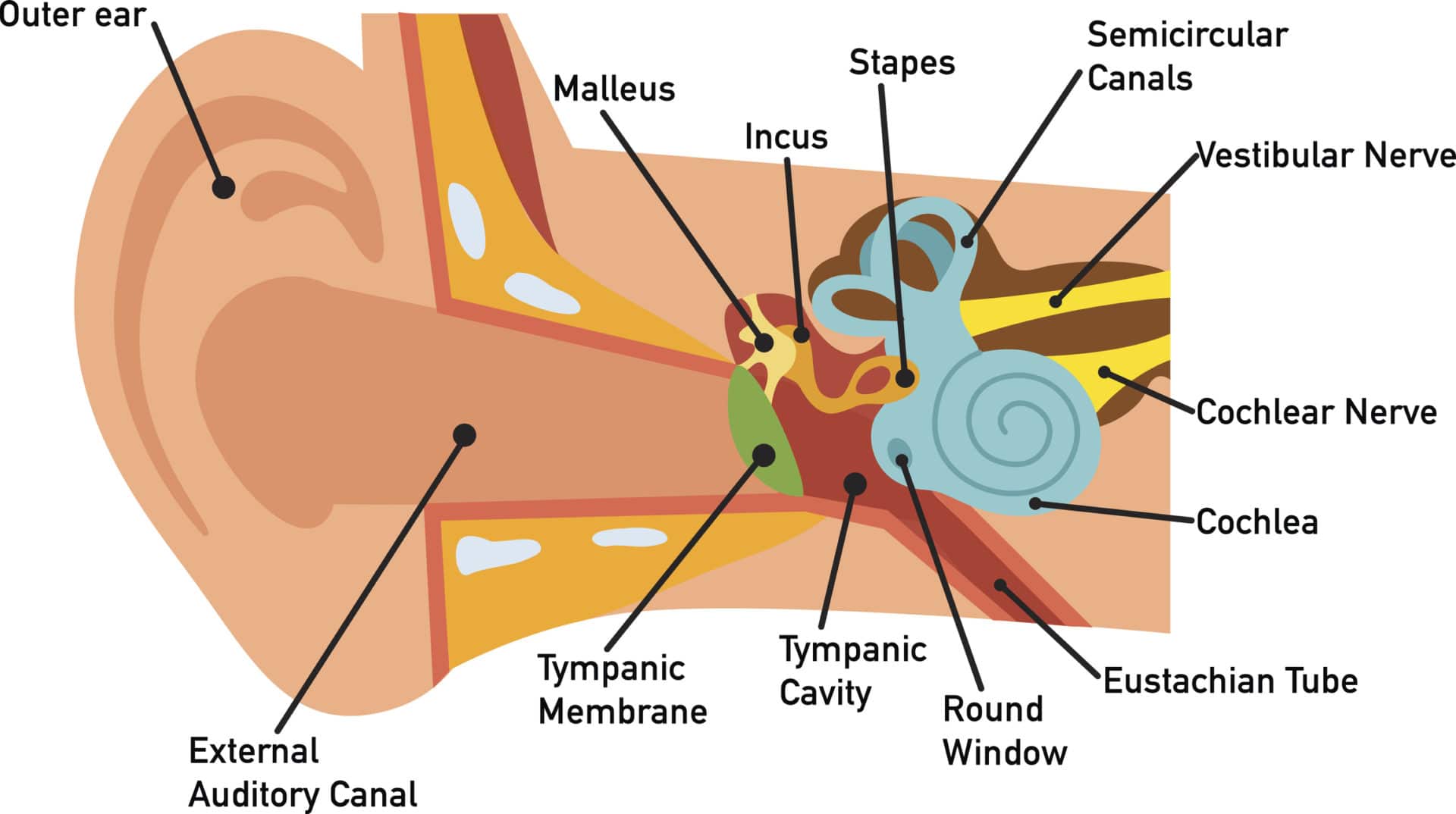
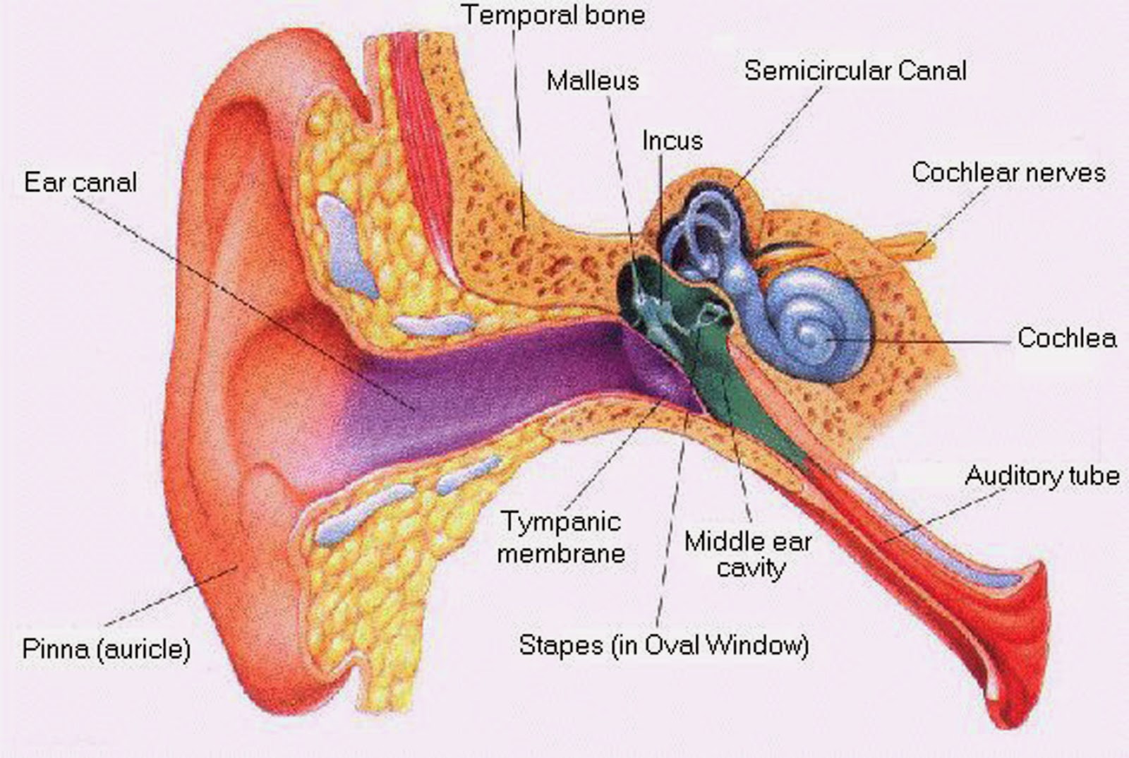
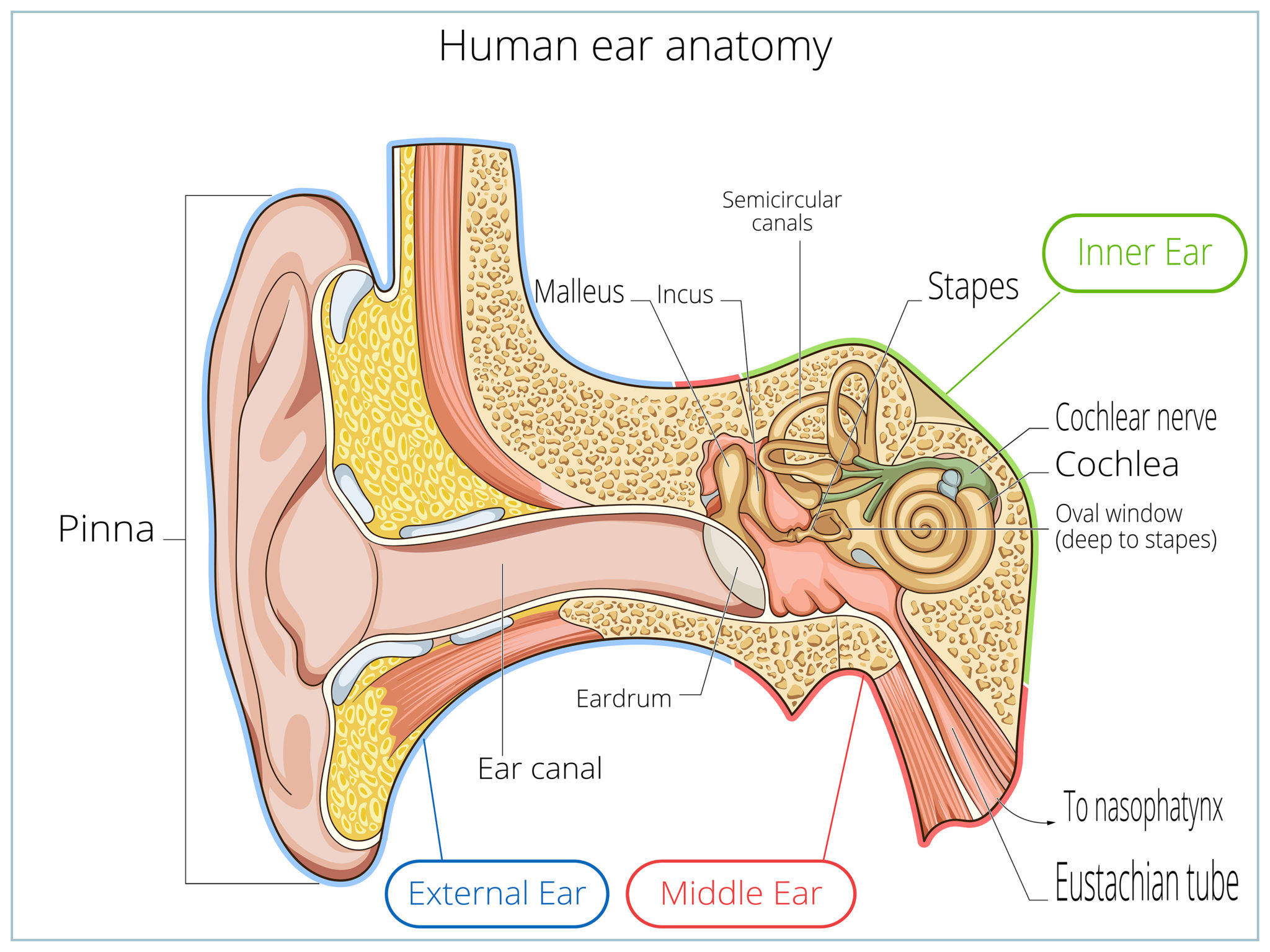
Anatomically, the ear has three distinguishable parts: the outer, middle, and inner ear. The outer ear consists of the visible portion called the auricle, or pinna, which projects from the side of the head, and the short external auditory canal, the inner end of which is closed by the tympanic membrane, commonly called the eardrum.
How You Hear Northland Audiology
Tympanogram Chapter 3 - Ear Anatomy Ear Anatomy - Outer Ear Ear Anatomy - Inner Ear Ear Anatomy Schematics Ear Anatomy Images Chapter 4 - Fluid in the ear Fluid in the ear Discussion Fluid in the ear Outline Middle Ear Ventilation Tubes Fluid in the ear Images Chapter 5 - Traveler's Ear Traveler's Ear Discussion Traveler's Ear Outline
30 Ear Diagram With Label Labels Design Ideas 2020
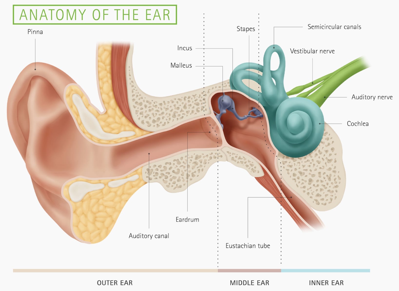
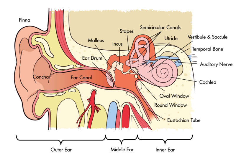
The ear is anatomically divided into three portions: External ear Middle ear Internal ear This mixture of bones, nerves, vessels, membranes, and muscles that make up the ear will be described in this article. Contents External ear Auricle External acoustic meatus Tympanic membrane Muscles of the external ear Vasculature of the external ear
Structure of the Ear Diagram Activity
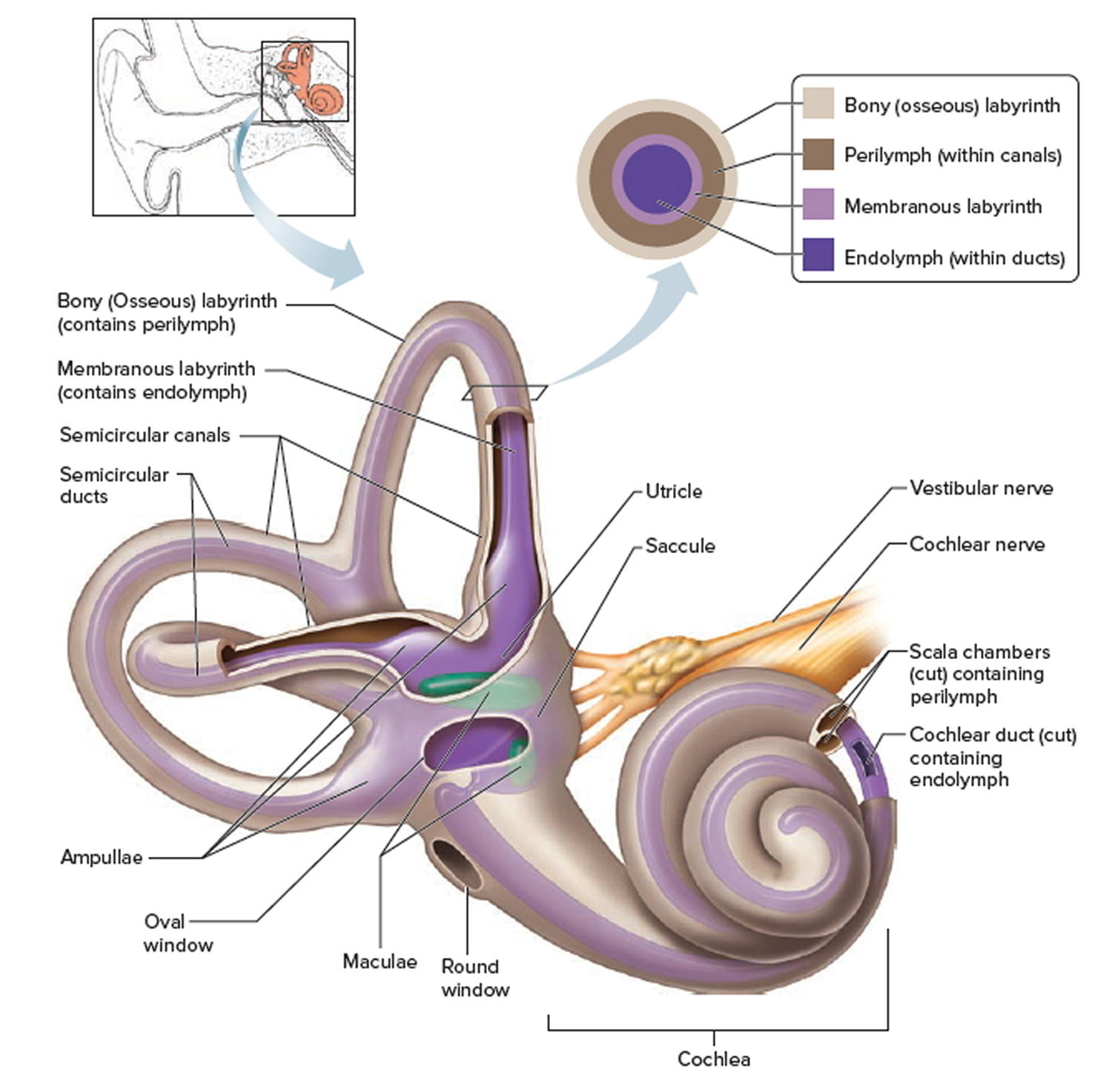
Your inner ear contains two main parts: the cochlea and the semicircular canals. Your cochlea is the hearing organ. This snail-shaped structure contains two fluid-filled chambers lined with tiny hairs. When sound enters, the fluid inside of your cochlea causes the tiny hairs to vibrate, sending electrical impulses to your brain.
Your Hearing Heritage Hearing
How to Draw Human Ear Diagram With Labelling #HumanEar - YouTube 0:00 / 5:50 How to Draw Human Ear Diagram With Labelling #HumanEar Articco Drawing 218K subscribers Subscribe Subscribed 8.3K.
HUMAN EAR OUTER EAR, MIDDLE EAR, INNER EAR, HEARING « SimpleBiology
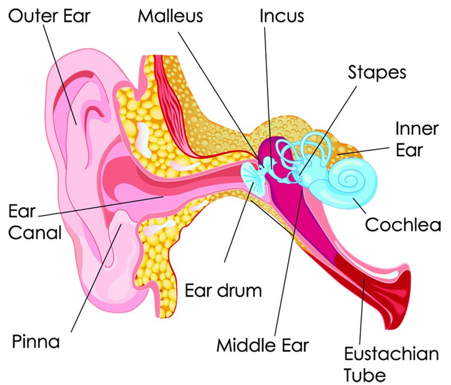
The anatomy of the ear consists of three main parts: the outer ear, middle ear and inner ear. This article will help explain each part to help you get a better understanding of the functions and anatomy of the ear. The Outer Ear
Hearing Noba
Diagram of Ear Human ear is a sense organ responsible for hearing and body balance. The outer ear receives the sound waves and transmits them down the ear canal to the eardrum. This causes the eardrum to vibrate and sound is produced. The diagram of the ear is important from Class 10 and 12 perspectives and is usually asked in the examinations.

How The Ear Works
The purpose of the inner ear is to sense and process information about sound and balance, and send that information to the brain. Each part of the inner ear has a specific function. Cochlea: The cochlea is responsible for hearing. It is made up of several layers, with the Organ of Corti at the center.
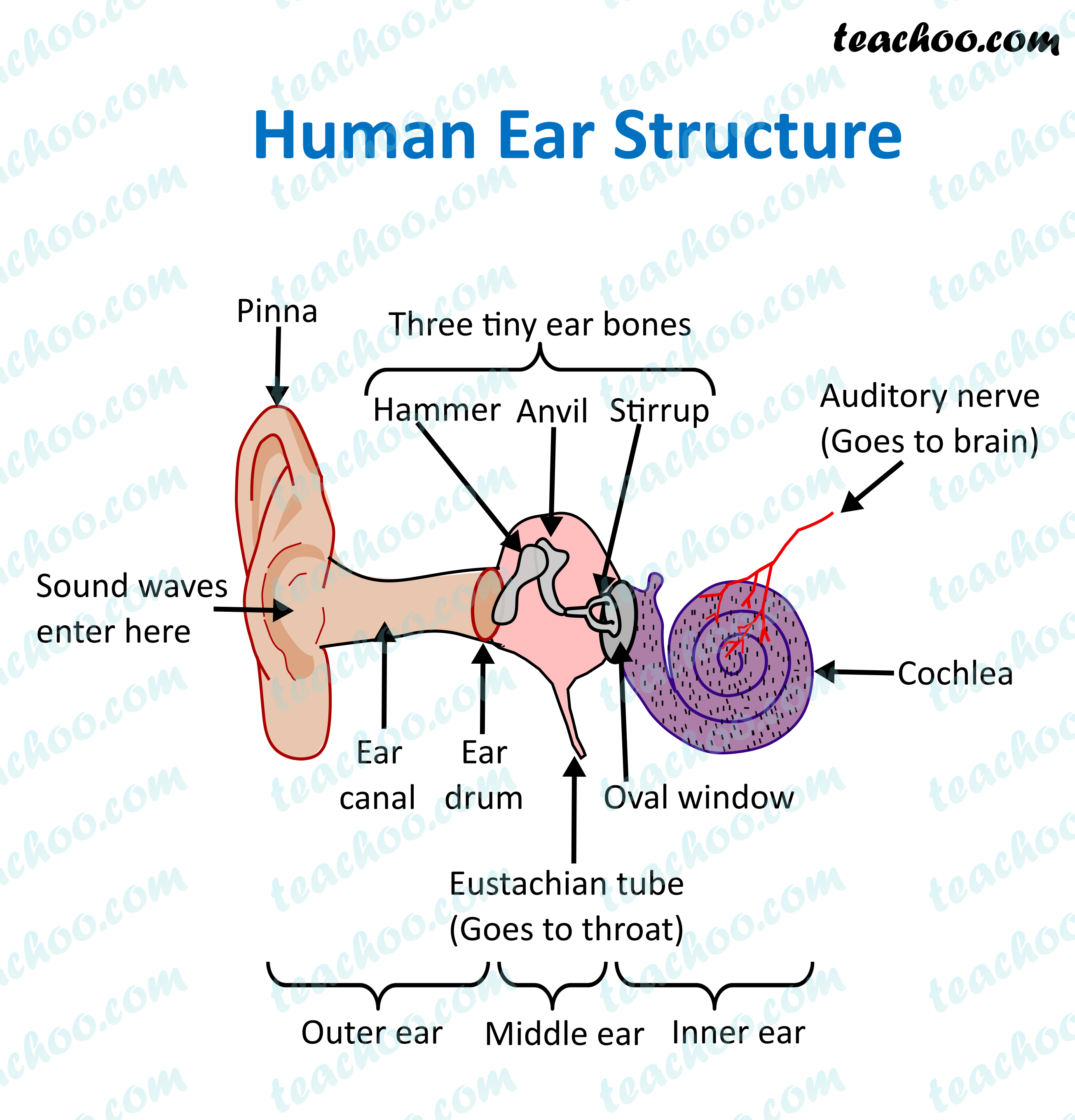
Structure and Function of Human Ear with Diagram Teachoo
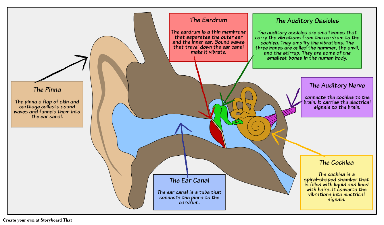
The tympanic membrane, or eardrum is the final hearing organ in the outer ear, separating it from the middle ear. The eardrum collects sound waves and vibrates, passing the sound waves into the middle ear. Most hearing disabilities are caused by trauma or disorders in the tympanic membrane eardrum.
The human ear structure and how it works Connect Hearing
Human ear. The ear is divided into three anatomical regions: the external ear, the middle ear, and the internal ear (Figure 2). The external ear is the visible portion of the ear, and it collects and directs sound waves to the eardrum. The middle ear is a chamber located within the petrous portion of the temporal bone.
Human Ear Anatomy Parts of Ear Structure, Diagram and Ear Problems
Get ready! Ear diagrams (labeled and unlabeled) Overview image showing the structures of the outer ear and auditory tube Take a moment to look at the ear model labeled above. This shows you all of the structures you've just learned about in the video, labeled on one diagram.
Ear Anatomy Causes of Hearing Loss Hearing Aids Audiology
The ear is divided into three parts: Outer ear: The outer ear includes an ear canal that is is lined with hairs and glands that secrete wax. This part of the ear provides protection and.
